Alpha Waves
Normal Occurrence of Alpha Waves
- Occipital Alpha is the dominant rhythm in relaxed wakefulness in 85% of healthy adults with closed eyes (Occipital = Occipital lobe at the back of the head).
- Alpha activity may spread to temporals and parietals in some people and this is considered normal.
- 85% of adults have a resting Alpha rhythm between 9.5-10.5 Hz.
- Some people have Alpha variants (slower or faster) that are generally considered normal if localized to the occipitals & attenuating (diminishing) with eye opening.
- Alpha may appear in polymorphic complexes mixed with Theta or Beta especially in drowsiness.
- Mu Rhythm (usually localized to C3 / C4 in scalp recorded EEG is considered an Alpha-like Rhythm & reflects motor system activation.
- Some individuals have an Alpha asymmetry over non-dominant hemisphere up to 50% greater and this is considered normal.
- Alpha activity generally attenuates (diminishes) with eye opening, mental activity, and drowsiness.
- Alpha activity may be present in REM sleep with a frontal / central distribution & may be related to periods of increased brain activity during sleep.
- Alpha may also be present with Delta in slow wave sleep
Abnormal Occurrence of Alpha Waves
- Frontally prominent & persistent Alpha may indicate hypometabolism (reduced blood flow) & abnormality.
- Alpha that fails to attenuate (diminish) with eye opening may be due to drowsiness or other pathology, including abnormalities in the visual system.
- Decreased Alpha peak frequency may reflect disease or brain injury such as TBI, dementia, medication effects, and age-related cognitive decline.
- Alpha peak frequency below about 8.5 Hz at PZ in the Adult waking record is considered slow.
- Alpha peak frequency above about 11 Hz at PZ in the Adult waking record is considered a fast variant.
- Absence of Alpha in one hemisphere or asymmetries between hemispheres greater than 50% are considered abnormal.
- Slowed Alpha rhythms may reflect neurological diseases such as dementia, Alzheimer’s, head injuries, or other conditions that can cause reduced cognitive abilities.
- Excessively fast Alpha can be associated with anxiety and OCD symptoms.
- Alpha that is frontally prominent may relate to depression and attention problems.
Physiological Origin of Alpha Wave Rhythms
- There are cortical generators of Alpha that act as “epicenters” where Alpha starts and spreads via cortico-cortical connections across the cortex.
- Alpha may also be generated by alternating GABAergic inhibition & Glutaminergic excitation of Thalamic “pace maker” neurons in the Reticular Nucleus.
- Cortical & Subcortical Alpha generators interact to cause the scalp recorded Alpha rhythm.
- Alpha amplitudes are influenced by cortical metabolism. Increased arousal usually = less Alpha.
- Alpha frequencies are influenced by the degree of Reticular Nucleus Neuronal Depolarization. More Activation / Depolarization = Faster Alpha Frequencies.
Role of Alpha Waves in Having a Healthy Brain
- The posterior Alpha rhythm is one of the best indicators of cognitive functioning.
- Alpha can be thought of as your brains “sampling rate” and indicates how your sensory system is interacting with your environment.
- Maintaining a healthy Alpha speed (@10 Hz for adults) is an important measure of cognitive functioning. If the Alpha begins to slow due to aging, head injury, toxic exposure, etc., cognitive problems can be expected.
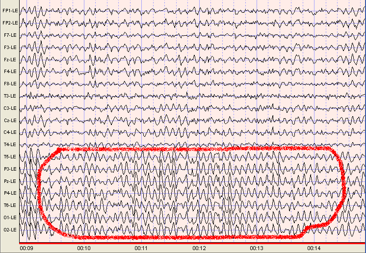
Images of Alpha Waves
Single channel;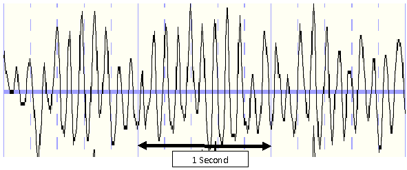
19 Channel: Linked Ear Montage;
40 year old female. Examples Highlighted in Red
Click here to learn about beta Waves >>>>>>>>>>>>>>>>>>>>>>>>
EEG Brainwaves
Alpha Waves
- Alpha Waves
- Normal Occurrence of Alpha Waves
- Abnormal Occurrence of Alpha Waves
- Physiological Origin of Alpha Wave Rhythms
- Role of Alpha Waves in Having a Healthy Brain
- Images of Alpha Waves