Reading Disabilities Treatment Program
Does your child have problems reading? If so, you are probably like many parents who can be frustrated watching their children struggle with their school work while other kids that seem no different to yours appear to doing it with ease. It may be a relief to know that you are not alone, it is suggested that approximately 15% of the population suffers with reading and/or spelling learning disabilities.
Here at the Scottsdale Neurofeedback Institute in Arizona we have been successfully treating learning disabilities for over 18 years. Our Reading Disabilities Treatment Program is based on a very successful and alternative treatment known as QEEG based EEG Neurofeedback.
A “learning disability” is defined as a 20 point gap between (IQ) intelligence and test scores. For instance with an IQ of 100 and reading at 80 or below. Another common definition of a learning disability is reading achievement scales two years below grade level.
Brain Mapping Research for Reading Disabilities
Neuroimaging research using individuals with a reading disorder has consistently found dysfunction in the left posterior temporal lobe (behind the left ear) and in the occipital lobe (visual cortex) in the back of the brain. We see letters in the visual cortex and attach sounds in the left posterior temporal lobe. If these areas are dysfunctional or disconnected or the timing is off, then reading/spelling is impaired. QEEG brain mapping indicates the types and severity of the dysfunction in this area and EEG neurofeedback can usually remediate the blood flow and metabolism abnormalities, timing, and connectivity dysfunctions in these areas.
QEEG Brain Maps and Discriminants for Reading Disabilities
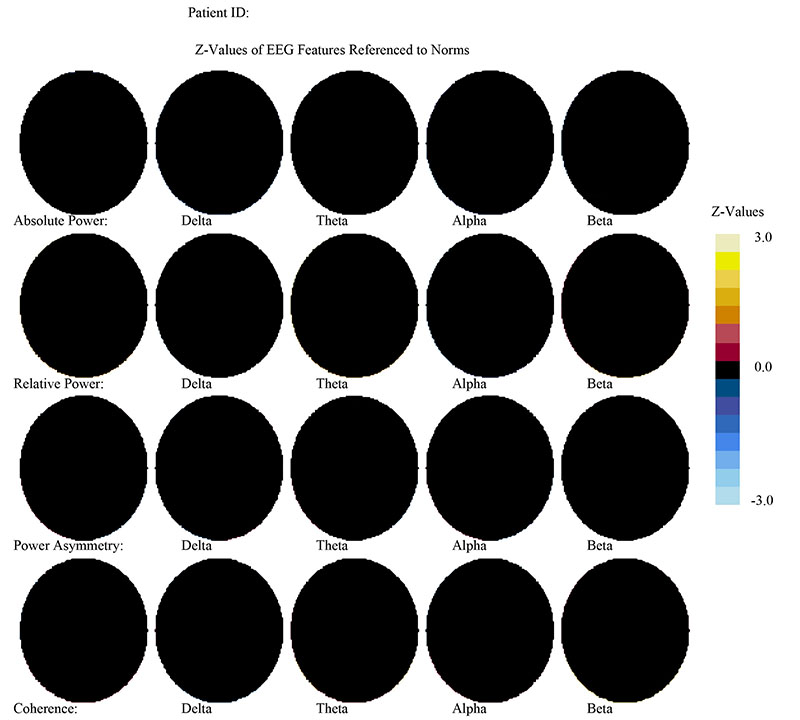
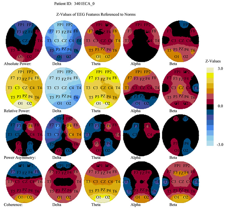
The images below are from a 7 year old female presenting with Attention Deficit Disorder, Oppositionality and reading difficulties. The map on the left is a representation of a normal working brain (all black) as produced by one of our six databases, the NX Link.
The map in the center is the actual brain map from our client. As mentioned previously, when a client presents with a reading disability, we expect to see elevated Delta, Theta, Alpha and/or Beta in the left posterior temporal lobe (behind the left ear) and in the occipital lobe (visual cortex) in the back of the brain. In the case of our client, abnormally elevated alpha at T5, P3, O1 and O2 is present in both relative and absolute power.
We also expect to see posterior (at the back of the head) coherence issues when a client presents with a reading disability, this again is present at the O1 – O2 regions.
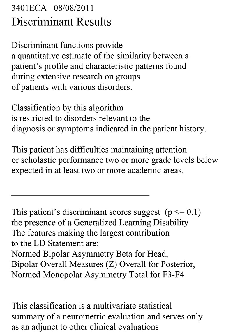
The image on the right is the Generalized Learning Disability Discriminant which is again produced by the NX Link database. The discriminant suggests the presence of a learning disability.


Neurofeedback, a primary treatment for Reading Disabilities
QEEG brain mapping, with multiple databases, including the New York University Medical School Normative Database, can accurately assess for what types of dysfunction are present and to what degree of severity. EEG neurofeedback training can, in most cases, rather remarkably remediate these abnormalities, with statistically significant improvements. With reading learning difficulties there is still usually a need for remediation such as tutoring, special education, computer and academic training to make up for the many years of underachievement caused by these areas performing inefficiently. Poor handwriting (Dysgraphia), located in the motor strip which runs from above one ear to the other, and facial expression processing deficits (such as Asperger’s) which is located in the fusiform gyrus, below the posterior temporal lobes are also treatable with neurofeedback. Pre and post mapping and pre and post testing is performed to measure improvement.For more information, or to schedule an appointment, call us on 480-625-4123
Contact
- 8114 E. Cactus Rd - Suite 200, Scottsdale, AZ, 85260
- +1 480 625 4123